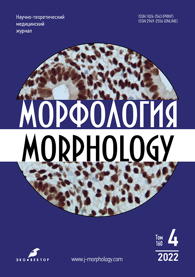
Vol 160, No 4 (2022)
- Year: 2022
- Published: 15.10.2022
- Articles: 5
- URL: https://j-morphology.com/1026-3543/issue/view/5575
- DOI: https://doi.org/10.17816/morph.20221604
Full Issue
Original Study Articles
Morphological changes in liver tissues of rats under the exposure of ecotoxicants and perinatal prophylaxis
Abstract
BACKGROUND: The high sensitivity of the liver to chemicals is due to its leading role in their metabolism. During the biotransformation of ecotoxicants, highly reactive intermediate products may be formed and free radical oxidation may be initiated, which may result in liver damage.
MATERIALS AND METHODS: The experiment was carried out on outbred white rats weighing 180–250 g. A total of 50 animals were involved in the experiment. All animals were divided into 5 groups: control and 4 experimental ones. All rats in the experimental groups were subjected to inhalation exposure to gasoline and formaldehyde vapors. In the 1st (control) group only poisoning with ecotoxicants was used, in the 2nd group peptinsorbent (apple pectin) was used against the background of poisoning with ecotoxicants, in the 3rd group — a membrane protector lemongrass, in the 4th group — a beets, in the 5th experimental group — peptinsorbent, membrane protector and beets. We examined pieces of liver and rats that were subjected to histological processing. Using a LEICA RM 2145 rotary microtome (Leica Microsystems, Germany), histological sections with a thickness of 5–8 μm were prepared. Stained sections were examined and photographed using an Axio Imager Z1 light microscope (Carl Zeiss, Germany).
RESULTS: The liver structure of rat pups born from female rats exposed to subchronic gasoline and formaldehyde poisoning throughout pregnancy has pronounced pathomorphological signs characteristic of hepatosis, turning into toxic hepatitis. The use of lemongrass, peptinsorbent and beets separately, along with intoxication of pregnant rats, somewhat reduced the degree of pathomorphological changes in the liver of born rat pups, but not dramatically. When using a combined mixture (apple pectin + membrane protector lemongrass + beets), the liver structure of the rat pups subsequently born was preserved relatively better than in the control group, with the exception of certain areas of the liver in which hemostasis and moderately pronounced dystrophic changes in hepatocytes were detected.
CONCLUSIONS: The combined mixture (apple pectin + membrane protector lemongrass + beets) has a more pronounced hepatoprotective effect compared to the separate use of each of its constituent substances and is effective as a hepatoprotective agent for liver damage from ecotoxicants.
 215-224


Morphological features of basal laminas of enteral nervous system structures in chronic slow transit constipation
Abstract
BACKGROUND: Studies on basal laminas in the tissues of the enteric nervous system are few and are carried out on experimental models in vivo and in vitro, performed on animals. The morphological features of the structure and localization of BL, their cellular sources of origin in various tissues of the gastrointestinal tract in normal and pathological conditions remain poorly studied.
AIM: Study of morphological features and distribution of basal laminas in human colon tissues and their changes in pathology.
MATERIALS AND METHODS: Fragments of the large intestine, obtained as a result of surgery for chronic slow-transit constipation, performed in S. M. Kirov Military Medical Academy were studied. The selective marker of basal laminas, type IV collagen, as well as neuronal and glial immunohistochemical markers (PGP 9.5, GFAP, S100â proteins) were used in the work
RESULTS: It has been shown that the greatest immunoreactivity within the intestinal wall is observed in the myenteric membrane, weak — in the vessels of the submucosa, locally expressed — in the subepithelial region of the mucous membrane (in the upper sections of the crypts). In smooth muscle, basal laminas were found around the smooth muscle cells of the longitudinal and concentric layers of the muscular membrane, mucous membrane, veins and arteries, as well as in the endothelium. It has been shown that the ganglionic Auerbach’s plexus is delimited from closely adjacent muscle layers of a continuous basal lamina, similar to the basal plate (glia limitans) of the brain and spinal cord of the CNS. It is clearly defined by its appearance — it has the form of a continuous hollow tubular structure. The sources of the formation of the basal plate around the ganglia of Auerbach and Meissner’s plexuses are various glial elements: in the first plexus, astrocyte-like and non-myelinated Schwann cells (both types located in the neuropil), and in the second plexus, glia of the autonomic nervous system (satellite cells of neurons and neurolemmocytes of postganglionic nerve fibers).
CONCLUSIONS: For the first time, the transition of basal lamina from the Auerbach’s plexus to numerous basal plates of neurolemmocytes of the Remakov fibers of the main terminal nerve plexus, which are involved in the innervation of the smooth muscle cells of the muscular membrane, is shown. Signs of dystrophic changes in the basal lamina structure associated with pathological changes in chronic slow transit constipation (tissue edema, inflammatory reactions, manifestations of agangliosis, gliosis, focal denervation of muscle cells, degeneration of nerve endings of neuromuscular and ganglionic plexuses) are shown in the work.
 225-238


Reviews
The features of the development and functioning of the kidneys in women in norm and pathology of pregnancy
Abstract
The kidneys play a central role for the emerging pregnancy, reacting and contributing to changes in homeostasis in the woman and fetus. Impaired kidney function during pregnancy is a fairly common and serious complication. Understanding the normal physiology of pregnancy provides the basis for further study of the changes that lead to impaired kidney function, and may provide the key to its better management.
This literature review systematizes information about the physiology and morphology of the kidneys before pregnancy, changes in them during its normal course and in cases of the presence or development of any pathological conditions for various reasons.
Knowledge of the anatomo-functional condition of the kidneys in normal pregnancy, as well as the changes that occur in renal diseases, will provide an opportunity to identify patients at high risk for the development of renal pathology, to define the management of pregnancy and, thereafter, to reduce the risk of maternal morbidity, mortality and perinatal losses.
 203-214


Biography
Sergey Vladimirovich Sazonov — to the 60-th anniversary of the birth
Abstract
The article is dedicated to the prominent Russian morphologist, Head of the Department of Histology of the Ural State Medical University, Honoured Worker of Higher Education of the Russian Federation, Doctor of Medical Sciences, Professor Sergey Vladimirovich Sazonov. On 17 May 2023 he turned 60 years old. Sergey Vladimirovich devoted his whole life to science, teaching, and his alma mater — Ural State Medical University. He opened a number of research and diagnostic laboratories in the Ural Federal District, continuously created and mastered new methods and new research technologies, introduced them into clinical medicine and education.
Sergey Vladimirovich was one of the organisers of the first scientific medical journal in our region — the Bulletin of the Ural State Medical Academy (now the Bulletin of Ural State Medical University). Sergey Vladimirovich successfully carried out material and technical re-equipment of the Department of Histology at Ural State Medical University, created a completely new in content multimedia course of lectures, and now he actively carries out digital transformation of his discipline, skilfully manages the creation of electronic educational resources in histology.
The article reflects the stages of life and creativity of Professor S. V. Sazonov, his main scientific achievements, professional and personal qualities.
 239-244


To the 125th anniversary of the birth of Ivan Nikitich Matochkin
Abstract
Ivan Nikitich Matochkin (1899–1973) is a prominent anatomist of the Soviet era, whose life story, as well as scientific and organizational work, should not be forgotten. The list of his merits should include the development of the department headed by him, participation in the organization of the Arkhangelsk branch of the All-Russian Scientific Society of Anatomists, Histologists and Embryologists, the Society «Knowledge», numerous scientific works on neuromorphology and angiology, the publication of scientific collections on research topics, management of Arkhangelsk State Medical Institute, defense of dissertations by many students of Professor Matochkin, which opened the way for them to a bright scientific future.
In this article, in addition to the main milestones in the life of Professor I. N. Matochkin, previously unknown archival data are presented that complement the pages of the biography of the scientist.
 245-249


