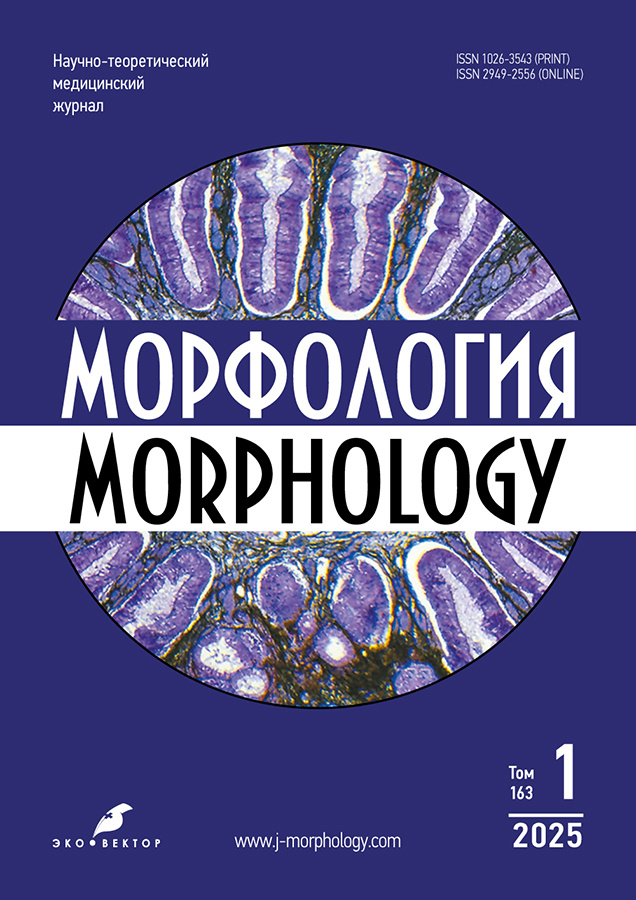
Vol 163, No 2 (2025)
- Year: 2025
- Published: 23.06.2025
- Articles: 8
- URL: https://j-morphology.com/1026-3543/issue/view/9883
- DOI: https://doi.org/10.17816/morph.20251632
Historical articles
Alexander Nikolaevich Bazhanov’s Contribution to the Development of Tissue Biology and Evolutionary Morphology
Abstract
The aim of this article is to analyze the contribution of Alexander Nikolaevich Bazhanov, a prominent Soviet histologist and representative of the Orenburg scientific histological school, to the development of fundamental problems in tissue biology and evolutionary morphology. Bazhanov was engaged in scientific research during the 1960s to 1990s. Bazhanov’s primary scientific research was devoted to the problems of embryonic histogenesis of the esophageal epithelium, the morphofunctional characteristics of esophageal mucosal structures, the study of evolutionary tissue changes, and tissue cultivation. His comparative histological studies and work on tissue culture led to the hypothesis that the esophageal epithelium originates from the prechordal plate. The findings obtained by Bazhanov served as the basis for the assertion that the prechordal plate is of endodermal origin. The results of his work made a substantial contribution to the development of the evolutionary direction in histology and promoted the advancement of research on debated problems in tissue biology. Bazhanov’s scientific legacy includes nearly one hundred articles and three monographs, two of which were co-authored. The importance of his work for the advancement of modern tissue biology was widely recognized during his lifetime. These studies have remained relevant even several decades after they were completed.
 83-92
83-92


Reviews
Structural and Functional Features of the Glymphatic System: a Contemporary Perspective
Abstract
The aging of the population in developed countries is a trend of major medical and social significance. In this regard, the study of the etiology and pathogenesis of neurodegenerative diseases, as well as the search for effective treatment methods, is of particular relevance. For a long time, it was believed that metabolic waste products were drained from the brain parenchyma’s interstitial fluid into the ventricular system. However, the discovery of the brain’s glymphatic system has significantly advanced our understanding of the mechanisms underlying pathologies associated with impaired clearance of metabolites from the brain. This scientific review outlines the main directions in the study of the functional morphology of the glymphatic system under normal conditions. It provides a detailed description of two theories of cerebrospinal fluid outflow and presents a critical analysis of both Russian and international research data. Under normal conditions, the function of the glymphatic system is influenced by heart rate, intracranial pressure, pulse and arterial pressure, as well as the phase of the respiratory cycle. In addition, sleep quality, head position during sleep, and exposure to toxic substances directly affect glymphatic system activity. The review also highlights recent data on the glymphatic system of the visual organs. Further research into the morphofunctional characteristics of the glymphatic system under normal conditions may greatly expand our fundamental understanding of disease pathogenesis and contribute to the development of new approaches to treatment and prevention.
 93-105


The Blood–Epididymis Barrier: Morphological, Physiological, Immunological and Seasonal Aspects and the Impact of Destabilizing Factors
Abstract
Spermatozoa entering the epididymis from the testis are unable to actively move and do not possess fertilizing ability. These functions are acquired within the lumen of the epididymal ducts, where the components of the blood–epididymis barrier create a specialized environment. The blood–epididymis barrier restricts paracellular transport and stimulates receptor-mediated transport of macromolecules across the epididymal epithelium. The blood–epididymis barrier consists of a pseudostratified columnar epithelium resting on a basement membrane, loose connective tissue of the lamina propria, and capillary endothelium located on its own basement membrane. Apical tight junctions and adherens junctions between adjacent principal cells of the pseudostratified epithelium play a key role in the blood–epididymis barrier’s function. Tight junctions are composed of various families of transmembrane proteins. The vascular component of the blood–epididymis barrier features continuous endothelium on an uninterrupted basement membrane. Alongside the epithelial and vascular components, interactions among dendritic cells, macrophages, and lymphocytes are critical in regulating blood–epididymis barrier permeability. In many species, the epididymis consists of 5 to 9 segments, each with distinct morphofunctional and biochemical characteristics. It has been shown that the barrier function becomes progressively more pronounced from the caput toward the cauda of the epididymis. Impaired function of intercellular junctions in the blood–epididymis barrier is considered a factor contributing to male infertility.
This review aimed to analyze the data on the morphofunctional organization of the blood–epididymis barrier.
 106-114


Original Study Articles
Histogenetic Features of Anorectal Lining Formation in Rats During Embryogenesis
Abstract
BACKGROUND: During embryonic development, the formation and differentiation of the mucosal tissues of the anal canal occur through the interaction of epithelia of different germ layer origins—ectodermal and enterodermal. The epithelial lining of the anorectal region of the rectum requires further characterization. Data on the histological structure and formation of tissue components in the anorectal canal during embryogenesis are relevant both theoretically (from an evolutionary perspective) and practically, as this region is a site of embryonic anomalies and neoplasms.
AIM: To describe the histogenetic features of anorectal lining formation in rats during embryogenesis.
METHODS: An observational, single-center, retrospective, uncontrolled study was conducted. The material consisted of the caudal portion of the anorectal canal of laboratory white rat (Rattus norvegicus) embryos at embryonic days 9, 13, 15, and 18. Morphological research methods were used. Histological analysis was performed on hematoxylin and eosin–stained sections.
RESULTS: On embryonic day 9, a cloacal membrane forms in the distal part of the embryo body. By day 13, the anorectal canal is formed and lined by cutaneous and intestinal epithelia, with a bilayered urothelium-like epithelium serving as a transitional interface. By day 15, this bilayered epithelium is replaced by clusters of rounded cells with hyperchromatic nuclei. On day 18, a clearly defined boundary is observed at the anorectal junction between the two genetically and morphologically distinct epithelia.
CONCLUSION: The findings indicate that, during embryogenesis in rats, a transient urothelium-like epithelium of the transitional zone exists between the cutaneous and intestinal segments of the anorectal canal, exhibiting distinct morphofunctional organization. In the later stages of embryogenesis, this transitional epithelium disappears, suggesting its provisional nature.
 115-122


Spatial Distribution Density of MyoD− and MyoD+ Nuclei in Muscle Fibers of Regenerating Skeletal Muscle Tissue: The Effect of Photobiomodulation
Abstract
BACKGROUND: Low-intensity laser exposure is used as a universal method for stimulating cellular activity, with photobiomodulatory effects directly dependent on laser wavelength and the absorption of radiation by specific tissue chromophores. It is known that infrared laser irradiation can stimulate cell proliferation and differentiation. The effects of green laser irradiation on tissue are poorly studied, and no studies have been found on the influence of low-intensity green photobiomodulation on cambial reserve cells of skeletal muscle. Meanwhile, the search for effective methods to restore skeletal muscle tissue after injury remains highly relevant.
AIM: To analyze the effects of infrared and green laser irradiation on the total number of nuclei in injured skeletal muscle fibers, as well as on the counts of MyoD+ (satellite cell nuclei) and MyoD− nuclei.
METHODS: The study was conducted on male Wistar rats divided into four experimental groups: Group 0, intact control (n = 8); Group I, rats with an incised skeletal muscle wound (n = 40); Group II, animals with an incised muscle wound treated with infrared laser irradiation (980 nm wavelength; n = 40); Group III, rats with an incised wound treated with green laser irradiation (520 nm wavelength; n = 40). Laser exposure was applied once in continuous mode for 180 seconds. The number of nuclei in the intact and injury zones of the skeletal muscle was assessed at 1, 3, 7, 14, and 30 days post-injury. Histological sections were stained with hematoxylin and eosin, as well as using immunohistochemistry with anti-MyoD antibodies.
RESULTS: The use of infrared and green photobiomodulation contributed to an increase in the spatial density of nuclei within skeletal muscle fibers in the injury zone on days 1 and 7 post-injury compared to animals in experimental Group I. Immunohistochemical analysis using anti-MyoD antibodies showed that the spatial density of MyoD+ and MyoD− nuclei increased in the injury zone on day 3 of the experiment compared to animals in Group I. Laser exposure resulted in an increased proportion of MyoD+ nuclei among the total nuclear population compared to animals in control Group I.
CONCLUSION: The application of infrared and green photobiomodulation promotes an early increase (on days 1 and 7) in the total number of nuclei in skeletal muscle fibers, with the effect of the 980 nm long-wavelength laser being more pronounced. In addition, the spatial density of MyoD+ satellite cells increased, with a more pronounced effect observed following green laser exposure. Activation of satellite cells by green laser light is early and short-term, whereas infrared irradiation induces a delayed but longer-lasting response. The overall findings indicate a stimulatory effect of laser exposure on regenerative processes in injured skeletal muscle.
 123-133


Morphological Features of the Kidneys in INSRR Knockout Mice Under Bicarbonate Load
Abstract
BACKGROUND: The insulin receptor-related receptor (IRR), a receptor tyrosine kinase, functions as a sensor of extracellular alkaline pH and is involved in renal bicarbonate excretion. High IRR expression has been detected in β-intercalated cells of the kidney, located in the distal tubules where bicarbonate secretion occurs. To create a new model for investigating sensitivity to pH changes, a unique INSRR knockout mouse line was developed on a C57BL/6 background.
AIM: To analyze morphological changes in kidney tissue in INSRR knockout mice compared with wild-type animals under normal conditions and during metabolic alkalosis.
METHODS: The study used littermate mice from a single generation, with genotypes confirmed by polymerase chain reaction. Two mouse lines were used in the experiment: INSRR knockout and wild-type. The animals were studied under two conditions—baseline and experimentally induced alkalosis. Morphometric analysis was performed on hematoxylin and eosin–stained kidney cryosections. The number of macrophages in kidney tissue was evaluated using immunohistochemical staining.
RESULTS: Morphometric analysis revealed that INSRR gene knockout did not lead to significant pathological alterations in kidney structure. However, significant differences were observed in parenchymal thickness, glomerular area, and collecting duct diameter in sections taken at the level of the renal pelvis. Differences were observed both when comparing the two mouse lines under normal conditions and during experimental alkalosis. Additionally, kidney size in knockout mice was smaller than in wild-type animals. Immunohistochemical analysis revealed no statistically significant differences in the number of CD206-positive (anti-inflammatory) macrophages in the kidneys under either normal conditions or experimental alkalosis.
CONCLUSION: Morphometric analysis of histological sections revealed increased parenchymal thickness in receptor tyrosine kinase knockout mice compared with wild-type animals under experimental alkalosis. Overall, knockout of the IRR receptor tyrosine kinase gene did not result in major pathological changes in kidney architecture. Thus, this genetically modified mouse line may serve as a model for physiological and molecular biological studies of metabolic alkalosis and its associated pathological processes.
 134-144


Somatometric and Bioimpedance Parameters in the Assessment of Physical Development in Patients with Esophageal Cancer
Abstract
BACKGROUND: Esophageal cancer is an oncological disease characterized by an extremely unfavorable course and prognosis. The high mortality rate associated with esophageal cancer underscores the importance of preventive screening programs in population and the need to expand the range of known risk factors for this disease. In this context, the study of physical development characteristics in patients with esophageal cancer is of particular interest.
AIM: To investigate somatometric and bioimpedance characteristics of physical development in patients with esophageal cancer.
METHODS: Anthropometric and bioimpedance measurements were conducted in 86 patients diagnosed with esophageal cancer. Body height and weight, shoulder and hip width, waist and hip circumference were measured. Body mass index and the sexual dimorphism index according to J.M. Tanner were calculated. Bioimpedance analysis was performed using the ABC01-036 hardware-software system (Medass, Russia). Body composition and phase angle were assessed. The phase angle of impedance reflects the intensity of metabolic processes in the body and is used as a prognostic indicator in oncological diseases. Statistical processing of the obtained data was performed using descriptive statistics methods. The Shapiro–Francia, Kolmogorov–Smirnov, Kruskal–Wallis, Fisher’s F-test, and Pearson’s chi-square test were used. Differences were considered statistically significant at p < 0.05.
RESULTS: Normal body weight was noted in most women with squamous cell carcinoma of the esophagus and in men with esophageal adenocarcinoma. Among male patients with esophageal cancer, the mesomorphic body type predominated, with a shift toward gynecomorphy (p = 0.001). A mesomorphic somatotype prevailed among female patients (p = 0.001). No statistically significant differences in bioimpedance parameters were found among patients with esophageal cancer depending on somatotype. Decreased phase angle values relative to the reference range established for the Russian population were recorded with equal frequency in patients with esophageal cancer, regardless of disease stage, which allows this parameter to be considered a potential marker of poor prognosis.
CONCLUSION: Body weight deficiency is not typical for patients with esophageal adenocarcinoma. Overall, patients with esophageal cancer tend to exhibit a mesomorphic somatotype, with a shift toward gynecomorphy in men, as well as decreased phase angle values regardless of disease stage.
 145-155


Biography
From the Heart to Science: 65th Anniversary of Professor Irina Alekseevna Odintsova
Abstract
On June 28, 2025, Professor Irina Alekseevna Odintsova—an esteemed histologist, talented educator, and outstanding individual—celebrates her 65th birthday. She is Head of the Department of Histology and Embryology at the S.M. Kirov Military Medical Academy, Doctor of Medical Sciences, Professor, and Honored Worker of Higher Education of the Russian Federation. This anniversary prompted us to briefly outline the key milestones of her biography and professional journey. Professor Odintsova’s life and work have been inextricably linked with the Military Medical Academy. For more than two decades, she has led the Department of Histology and Embryology, where her students and colleagues work productively and gain scientific experience under her guidance. Professor Odintsova is engaged not only in scientific research but also in extensive educational outreach. She initiated the tradition of organizing national histological conferences and academic meetings not only within the department but also at prominent venues across Saint Petersburg, transforming these events into noteworthy and anticipated gatherings for the entire Russian histological community. Her students, colleagues, and friends extend their warmest congratulations to Irina Alekseevna, wishing her good health, professional longevity, boundless energy, and happiness.
 156-161


