Морфология
Рецензируемое научно-теоретическое медицинское периодическое издание.
Главный редактор
- Деев Роман Вадимович
кандидат медицинский наук, первый заместитель директора
НИИ Морфологии человека им. акад. А.П. Авцына ФГБНУ РНЦХ им. акад. Б.В. Петровского
ORCID iD: 0000-0001-8389-3841
Издатель
- Эко-Вектор (https://eco-vector.com/)
Учредители
О журнале
Журнал «Морфология» (ранее - "Архив анатомии, гистологии и эмбриологии") является ведущим морфологическим научным журналом России, который выпускается непрерывно с 1916 г.
Журнал был основан крупным отечественным гистологом А.С. Догелем, в составе его редколлегии на протяжении многих лет работали наиболее выдающиеся отечественные ученые, передавшие свою эстафету современному поколению морфологов.
ВАК
Журнал включен в перечень рецензируемых научных изданий, в которых должны быть опубликованы основные научные результаты диссертаций на соискание ученой степени кандидата наук, на соискание ученой степени доктора наук.
В соответствии с Постановлением Правительства РФ от 24.09.2013 N 842 (в редакции от 11.09.2021) "О порядке присуждения ученых степеней" (вместе с "Положением о присуждении ученых степеней") и Постановлением Правительства РФ от 20.03.2021 N 426 "О внесении изменений в некоторые акты Правительства Российской Федерации и признании утратившим силу постановления Правительства Российской Федерации от 26 мая 2020 г. N 751":
... п. 11. Основные научные результаты диссертации должны быть опубликованы в рецензируемых научных изданиях (далее - рецензируемые издания). К публикациям, в которых излагаются основные научные результаты диссертации, в рецензируемых изданиях приравниваются публикации в научных изданиях, индексируемых в международных базах данных Web of Science и Scopus и международных базах данных, определяемых в соответствии с рекомендацией Комиссии (далее - международные базы данных), а также в научных изданиях, индексируемых в наукометрической базе данных Russian Science Citation Index (RSCI). ...
Тематики публикуемых статей
(в соответствии с новой номенклатурой специальностей ВАК)
- 1.5.22. Клеточная биология (биологические науки*, медицинские науки)
- 1.5.23. Биология развития, эмбриология (биологические науки, медицинские науки)
- 3.3.1. Анатомия человека (медицинские науки)
- 3.3.2. Патологическая анатомия (медицинские науки, биологические науки)
- 4.2.1. Патология животных, морфология, физиология, фармакология и токсикология (ветеринарные науки, биологические науки)*
Типы принимаемых к рассмотрению рукописей
- Научные обзоры
- Систематические обзоры и метаанализы
- Результаты оригинальных исследований
- Клинические случаи и серии клинических случаев
- Краткие сообщения
- Письма в редакцию
- Клинические рекомендации
Публикации
- на русском и английском языке;
- в составе регулярных выпусков каждый квартал (4 раза в год);
- на сайте журнала в режиме Online First по мере принятия произведений к публикации;
- бесплатно для авторов;
- в гибридном доступе - по подписке и открыто
(статьи в Open Access распространяются на условиях открытой лицензии Creative Commons-NonCommertical-NonDerivates-Attribution 4.0 International (CC BY-NC-ND 4.0)).
Индексация
- Russian Science Citation Index (RSCI)
- Ядро РИНЦ (Российский индекс научного цитирования)
- Белый список
- ВАК К2
- НЭБ (eLibrary.ru)
- Google Scholar
- Ulrich's Periodicals Directory
- Dimensions
- Crossref
- Scilit
- OpenAlex
- Wikidata
- Scholia
- Fatcat
Объявления Ещё объявления...

Уникальная скидка на подписку до 1 декабря 2025 года!Размещено: 24.10.2025
Уважаемые коллеги! Активным читателям издательство дарит скидку 20% на приобретение подписного абонемента на 2026 год. Вам доступна возможность оформления подписки на журналы в бумажном варианте или цифровой форме. Вы можете оформить необходимый журнал непосредственно на нашем веб-сайте или подать заявление на электронный почтовый ящик: podpiska@eco-vector.com Специальное предложение действительно для физических лиц до 1 декабря 2025 года! |
|
|

Журнал «Морфология» включен в «Белый список» (ЕГПНИ)Размещено: 10.10.2025
Дорогие коллеги! С радостью сообщаем вам, что журнал «Морфология» включен в «Белый список» — Единый государственный перечень научных журналов (ЕГПНИ). По итогам категорирования журналу присвоен У2. URL: https://journalrank.rcsi.science/ru/record-sources/details/29798/ В соответствии с Постановлением Правительства РФ №1494 от 06.11.2024, опубликованные в нашем журнале статьи могут быть использованы для оценки публикационной активности авторов и представляемых ими организаций. |
|
|

Подписная кампания 2026 года стартовала: специальное предложение!Размещено: 12.09.2025
1 сентября открыта подписная кампания 2026 года. Издательство «Эко-Вектор» сохраняет цены для самых активных пользователей*, успей подписаться на новый год по цене старого! Для читателей – это отличный шанс получить годовую подписку на печатные версии журналов по ценам 2025 года! Предложение действует до 1 декабря 2025 года! *только для физических лиц |
|
|
Текущий выпуск
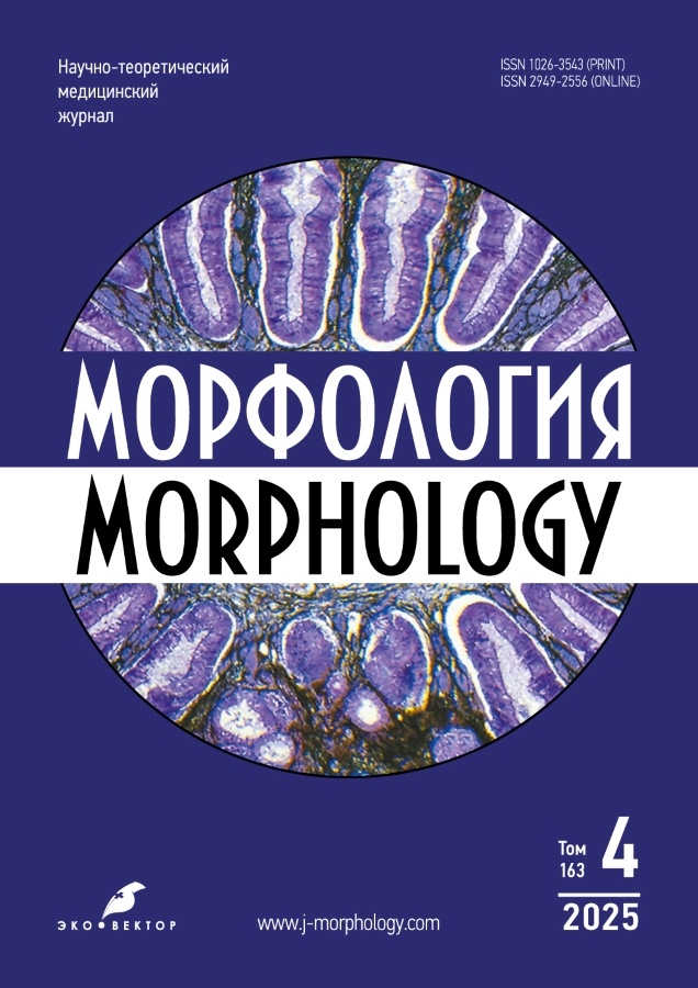
Том 163, № 4 (2025)
- Год: 2025
- Выпуск опубликован: 23.10.2025
- Статей: 12
- URL: https://j-morphology.com/1026-3543/issue/view/9885
- DOI: https://doi.org/10.17816/morph.20251634
Исторические статьи
Известный отечественный морфолог Ираида Михайловна Пестова
Аннотация
Статья посвящена памяти Ираиды Михайловны Пестовой (1913–1988) — видного отечественного гистолога советского периода, профессора, доктора биологических наук. Ираида Михайловна была выпускницей Свердловского государственного медицинского института (ныне Уральский государственный медицинский университет), представительницей его легендарного первого выпуска. По окончании института в 1936 году И.М. Пестова поступила в аспирантуру на кафедру нормальной анатомии, где в течение года занималась под руководством профессора Алексея Павловича Лаврентьева — основателя и первого заведующего этой кафедрой. В 1937 году Ираида Михайловна переехала в город Пермь, где была зачислена в аспирантуру при кафедре гистологии Пермского государственного медицинского института, к профессору Петру Яковлевичу Лаховскому. С этого момента начался её успешный путь учёного-гистолога, приведший к выдающимся научным достижениям и многолетнему заведованию кафедрой гистологии Пермского государственного медицинского института. Главным научным направлением кафедры в тот период было изучение иммунной и кроветворной систем позвоночных животных в эволюционном аспекте. Как учёный, Ираида Михайловна Пестова была последовательницей выдающегося советского гистолога, основоположника эволюционной гистологии, академика Академии Наук и Академии Медицинских Наук СССР Алексея Алексеевича Заварзина. Именно в Перми А.А. Заварзин заложил научные основы эволюционного направления в гистологии, которые в дальнейшем плодотворно развивал Евгений Сильвиевич Данини — учитель И.М. Пестовой. В статье отражены основные этапы жизни и деятельности профессора И.М. Пестовой, её вклад в науку и преподавание, а также профессиональные и личные качества.
 256-264


Научные обзоры
Применение автоматизированных систем для создания тканевых микроматриц в онкоморфологических исследованиях
Аннотация
Тканевые микроматрицы — один из перспективных методов для высокопроизводительного анализа архивированных образцов тканей. Лаборатории, использующие традиционный ручной метод изготовления тканевых микроматриц (ТМА), сталкиваются с необходимостью повышения эффективности и стандартизации, что особенно важно для онкоморфологических исследований и диагностики. Достичь этого можно за счёт автоматизации процесса.
Настоящий обзор посвящён рассмотрению возможностей и преимуществ автоматизированных систем для создания ТМА по сравнению с ручным методом, с акцентом на их применение в анализе саркомы Юинга и других недифференцированных круглоклеточных сарком. В проанализированных работах автоматизированные системы использовали для извлечения и позиционирования тканевых цилиндров в парафиновые блоки-реципиенты, а на полученных срезах ТМА проводили гистологические и иммуногистохимические исследования. Кроме того, оценивали качество срезов тканевых микроматриц. В упомянутых работах автоматизированные системы показали высокую точность позиционирования тканевых цилиндров, что значительно ускорило процесс создания ТМА и улучшило качество готовых срезов.
Таким образом, внедрение автоматизированных систем для конструирования ТМА имеет значительные преимущества по сравнению с ручным методом, поскольку обеспечивает стандартизацию и повышает производительность лабораторных исследований. Автоматизированные системы позволяют эффективно анализировать большие серии образцов, что особенно важно для валидизации диагностических и прогностических биомаркеров. Опубликованные работы подчёркивают необходимость дальнейшего развития и более широкого внедрения автоматизированных систем в онкоморфологические исследования для повышения их эффективности и воспроизводимости.
 265-272


Методологические аспекты применения искусственного интеллекта для морфологической диагностики фиброза, дистрофии и воспалительных поражений печени
Аннотация
Неопухолевые заболевания печени широко распространены и остаются сложными для диагностики. По современным данным распространённость неалкогольной жировой болезни печени среди взрослых в России составляет около 25%. Морфологическая верификация фиброза, жировой и баллонной дистрофии, воспалительной инфильтрации и некроза ткани печени, зависит от субъективного мнения специалиста, что затрудняет стандартизацию. По этим причинам разработка объективных и автоматизированных методов анализа морфологических изменений в печени может значительно повысить воспроизводимость диагностики. В данном обзоре проведён анализ современных подходов к применению методов искусственного интеллекта в морфологической диагностике неопухолевых поражений печени, а также рассмотрены основные направления использования нейросетевых алгоритмов, включая классификацию и сегментацию гистологических изображений. Кроме того, проведена оценка эффективности разработанных моделей при выявлении основных морфологических паттернов: фиброза, баллонной и жировой дистрофии, воспалительной инфильтрации.
Для обзора были использованы публикации, найденные в базах данных Google Академия и PubMed. Поиск охватывал период с 2020 по 2025 год, в окончательный анализ включены 22 публикации.
Установлено, что модели искусственного интеллекта демонстрируют высокую точность, которая, однако, зависит от объёма выборки, учёта межлабораторной вариабельности, морфологического паттерна, выбора увеличения микроскопа и метода окрашивания микропрепаратов. Для дальнейшего развития данного направления требуется увеличение объёма открытых данных и стандартизация подходов. Тем не менее, модели разрабатываются даже на небольших объёмах данных, что делает методику доступной для широкой исследовательской аудитории.
 273-282


Оригинальные исследования
Количественная характеристика популяции тучных клеток селезёнки лабораторных мышей при экспериментальном облучении рентгеновским излучением
Аннотация
Обоснование. Количественная и морфофункциональная характеристики тучных клеток могут служить одним из показателей реактивности тканей в ответ на радиационное воздействие, а также критерием компенсаторно-приспособительных процессов после облучения и при использовании радиопротекторов.
Цель — представить морфофункциональную и количественную характеристики тучных клеток селезёнки лабораторных мышей при фракционированном общем рентгеновском облучении и пероральном введении бета-D-глюкана.
Методы. Проведено экспериментальное одноцентровое проспективное сплошное контролируемое исследование. Объект исследования — образцы селезёнки лабораторных мышей (n = 23). Количественно оценивали популяцию тучных клеток на гистологических срезах селезёнки. Мышей разделили на 5 групп: 1 — интактные животные (n = 3); 2 — облучённые животные с суммарной поглощённой дозой 7 Гр (n = 5); 3 — облучённые мыши с суммарной поглощённой дозой 7 Гр, которым перорально вводили растворимую форму бета-D-глюкана за 15 мин до облучения (n = 5); 4 — облучённые животные с суммарной поглощённой дозой 18 Гр (n = 5); 5 — облучённые мыши с суммарной поглощённой дозой 18 Гр, которым перорально вводили растворимую форму бета-D-глюкана за 15 мин до облучения (n = 5). Взятие материала осуществляли на 14 и 30 сутки после начала экспериментального воздействия. Образцы фиксировали в 10% растворе забуференного формалина, обезвоживали в спиртах и заливали в парафин. Срезы окрашивали по методу Романовского–Гимзы. На каждом гистологическом препарате оценивали структуру и подсчитывали количество тучных клеток. Проводили статистическую обработку полученных данных.
Результаты. Плотность расположения тучных клеток в селезёнке лабораторных мышей при поглощённой суммарной дозе облучения 7 Гр изменилась незначительно по сравнению с интактными животными. При суммарной поглощённой дозе 18 Гр отмечено значительное увеличение плотности расположения и функциональной активности тучных клеток. Предварительное введение бета-D-глюкана перед облучением в суммарной поглощённой дозе 7 Гр снижает количество тучных клеток в 2,5 раза, а при суммарной дозе 18 Гр — в 1,25 раза по сравнению с облучёнными животными без введения препарата (группа сравнения 4).
Заключение. Плотность расположения тучных клеток в селезёнке зависит от поглощённой дозы рентгеновского излучения. Введение бета-D-глюкана за 15 мин до воздействия снижает плотность расположения тучных клеток, что, вероятно, можно рассматривать как положительный радиопротекторный эффект.
 283-292


Ультраструктурные изменения гломерулярного фильтрационного барьера почек у крыс при остром отравлении параоксоном
Аннотация
Обоснование. Параоксон — фосфорорганическое соединение, хроническое отравление которым имеет различные проявления. У лиц, контактирующих с фосфорорганическими соединениями, нередко выявляются гломерулярный или тубулярный склероз, а также их сочетание. Описание ультраструктурных изменений гломерулярного фильтрационного барьера (ГФБ) почек при экспериментальном моделировании острого отравления параоксоном на данный момент в доступной литературе отсутствует, что подчёркивает актуальность данного исследования.
Цель — выявить ультраструктурные изменения гломерулярного фильтрационного барьера почек у крыс при остром отравлении сублетальными дозами параоксона.
Методы. Фрагменты почек получены от самцов белых беспородных крыс Rattus norvegicus спустя 1, 3 и 7 суток после отравления параоксоном. В работе использовали три режима воздействия параоксона. Проведены иммуногистохимическое, электронно-микроскопическое и морфометрическое исследования почек, а также статистический анализ результатов.
Результаты. Ультраструктурные изменения ГФБ почек наблюдаются во всех режимах воздействия параоксона. В эндотелиальных клетках клубочковых капилляров почечных телец увеличивается диаметр фенестр при одновременном уменьшении частоты их расположения. В подоцитах обнаружено изменение размеров подошвенной части цитоподий и сокращение количества отростков III порядка, примыкающих к гломерулярной базальной мембране. У животных всех трёх опытных групп увеличивается толщина гломерулярной базальной мембраны на 7 сутки после острого отравления. Наиболее выраженные морфологические изменения структур ГФБ после воздействия параоксона наблюдали в эндотелиальных клетках клубочковых капилляров.
Заключение. Острое отравление параоксоном вызывает ультраструктурные изменения ГФБ почек у крыс. Полученные данные способствуют пониманию механизмов развития гломерулосклероза при хроническом воздействии токсиканта.
 293-304


Морфометрические особенности интрамуральных автономных нервных ганглиев межмышечного и подслизистого сплетений тонкой и толстой кишки крыс в постнатальном онтогенезе
Аннотация
Обоснование. Морфология интрамуральных автономных нервных ганглиев межмышечного (МС) и подслизистого (ПС) сплетений кишки у половозрелых животных изучена достаточно подробно, тогда как данных о возрастных особенностях этих структур в современной литературе недостаточно.
Цель — исследовать морфометрические характеристики интрамуральных автономных нервных узлов межмышечного и подслизистого сплетений в тонкой и толстой кишке у крыс в постнатальном онтогенезе.
Методы. Работа выполнена на самцах крыс линии Wistar разных возрастных групп: новорождённых, на 10, 20, 30, 60-е сутки после рождения, а также в возрасте 12 и 24 месяца. В работе использовали иммуногистохимический анализ с флуоресцентными меченными антителами к протеиновому генному продукту 9,5 (PGP9.5).
Результаты. В постнатальном онтогенезе происходит снижение числа ганглиев на 1 мм2 и увеличение площади ганглиев в тонкой и толстой кишке. Средняя площадь нервных узлов в МС тонкой и толстой кишки возрастает с момента рождения вплоть до 60-х суток, а в ПС — в первые 30 суток жизни. Средняя плотность расположения нервных узлов на 1 мм2 уменьшается в МС: в тонкой кишке в первые 60 суток, а в толстой — на протяжении 12 месяцев. Данный показатель в ПС снижается и в тонкой, и в толстой кишке в первые 60 суток жизни. Среднее число PGP9.5-иммунореактивных нейронов в одном ганглии в МС не изменяется в постнатальном онтогенезе, а в ПС увеличивается в первые 10 суток после рождения.
Заключение. В постнатальном онтогенезе в первые 30 суток жизни происходит увеличение размеров нервных узлов в МС и ПС и снижение плотности их расположения на единицу поверхности тонкой и толстой кишки. Форма ганглиев и число нейронов в узлах МС в постнатальном онтогенезе не меняется. Ганглии ПС, в отличие от МС тонкой и толстой кишки крыс, к моменту рождения остаются незрелыми и формирование сети узлов ПС происходит в первые 10 суток после рождения.
 305-315


Особенности эпиморфной регенерации дистальной фаланги пальца у иглистых мышей (Acomys cahirinus)
Аннотация
Обоснование. Восстановление концевой фаланги пальца у взрослых млекопитающих представляет собой редкий пример полноценной регенерации без развития фиброза. Иглистые мыши рода Acomys способны к восстановлению многих тканей, включая эпидермис и мышцы, без формирования рубца. Однако способность Acomys cahirinus к полноценному восстановлению концевой фаланги пальца не известна.
Цель — оценить динамику восстановления концевой фаланги пальца после ампутации у иглистых мышей.
Методы. Динамику регенерации концевых фаланг пальцев у мышей Mus musculus и A. cahirinus оценивали в течение 28 дней после ампутации. Для моделирования эпиморфной регенерации посредством формирования бластемы ампутацию концевой фаланги проводили дистальнее ногтевого ложа. Для моделирования повреждения, завершающегося формированием фиброзного рубца, ампутацию проводили проксимальнее ногтевого ложа. Восстановление оценивали визуально, с использованием микрокомпьютерной томографии, а также на основании результатов гистологического исследования.
Результаты. В отличие от M. musculus, демонстрирующих полноценное восстановление всего тканевого состава после ампутации дистальной фаланги пальца, у A. cahirinus этого не происходит. У A. cahirinus после ампутации дистальнее ногтевого ложа наблюдается укорочение пальцев и их деформация по типу «барабанных палочек». Гистологический анализ показал, что у иглистых мышей увеличивается объём костной ткани в повреждённой фаланге. С помощью микрокомпьютерной томографии установлено, что повреждённая при ампутации кость подвергается лизису вплоть до полной деградации, а гипертрофия происходит в кости следующей фаланги повреждённого пальца, расположенной проксимальнее плоскости ампутации.
Заключение. У A. сahirinus не происходит полноценного восстановления пальца после ампутации дистальной фаланги. Возможно, это обусловлено чрезмерным лизисом повреждённой кости, а также недостаточным формированием бластемы в участке повреждения.
 316-326


Соотношение пролиферации и апоптоза в клетках соединительных тканей кожи при заживлении механической травмы в эксперименте
Аннотация
Обоснование. Важным аспектом течения раневого процесса являются процессы пролиферации и апоптоза в зоне повреждения. Особый интерес представляет характеристика регенерационного гистогенеза тканей кожи в перинекротической области раны, а именно соединительнотканные слои — дерма и гиподермис. Специфика перинекротической области состоит в том, что в ней располагаются камбиальные элементы эпителиальных и соединительных тканей, за счёт которых осуществляется регенерация, а процессы клеточной гибели имеют характерные особенности. Для изучения закономерностей гистогенетических процессов, в том числе пролиферации и клеточной гибели, в тканях с различной камбиальностью чаще всего используют иммуногистохимические методы. Однако по-прежнему сохраняет свою актуальность проблема выбора маркеров, отражающих соотношение процессов пролиферации и апоптоза на разных этапах регенерации.
Цель — провести иммуногистохимическую оценку соотношения пролиферации и апоптотической гибели клеток соединительных тканей кожи на разных этапах заживления механической раны.
Методы. Проведено экспериментальное одноцентровое сплошное контролируемое рандомизированное неослеплённое исследование. Материалом служили образцы кожи бедра крыс линии Wistar на разных этапах заживления после механического повреждения (нанесения глубокой резаной раны). Животных разделили на 9 групп: контрольная группа — интактные крысы (n = 3); остальные группы соответствуют срокам после нанесения механической травмы — 12 ч, 24 ч, 2, 3, 6, 10, 15 и 25 суток (n = 3 в каждой группе). Из фрагментов кожи готовили препараты для гистологического и иммуногистохимического исследования. Для выявления процессов пролиферации использовали антитела к фосфорилированному гистону Н3, для выявления апоптоза — антитела к белку р53 и каспазе 3.
Результаты. В соединительных тканях кожи крыс всех экспериментальных групп обнаружены иммунопозитивные клетки, экспрессирующие фосфогистон Н3, каспазу 3 и белок р53. Определён индекс пролиферации клеток и проанализирована динамика экспрессии проапоптотических белков в интактной коже и в перинекротической области на разных этапах процесса регенерации. На основании полученных данных были рассчитаны пролиферативно-апоптотическое соотношение и числовой показатель (индекс), характеризующий оба процесса. Показано, что пролиферативно-апоптотический индекс достигает наиболее высоких значений в периоды преобладания пролиферации над процессами клеточной гибели — в интактной коже и на завершающих этапах регенерации, минимальные значения индекс принимает при преобладании процессов апоптоза — в фазу воспаления и некроза.
Заключение. В работе впервые использована иммуногистохимическая реакция с антителами к фосфогистону Н3 для исследования процесса регенерации кожной раны. Этот маркер экспрессируется пролиферирующими клетками и в совокупности с маркерами апоптоза позволяет определить пролиферативно-апоптотическое соотношение на разных этапах регенерации.
 327-338


Краниоскопические и краниометрические характеристики мозгового черепа взрослого человека в норме и при деформациях
Аннотация
Обоснование. Современные краниологические исследования должны основываться на комплексном подходе и включать алгоритмы отделения нормы от патологии. Важно знать, насколько краниометрические параметры черепов, относящихся к деформированным, отклоняются от нормы.
Цель — изучить краниоскопические и краниометрические характеристики мозгового черепа в норме и при различных деформациях на основе материалов коллекции Б.А. Долго-Сабурова, хранящейся в фундаментальном музее кафедры нормальной анатомии Военно-медицинской академии; определить частоту встречаемости деформаций, а также разработать критерии дизайна современного краниологического исследования.
Методы. Исследована серия из 842 черепов. Оценивали состояние швов, выявляли признаки краниосиностозирования и наличия сопутствующих аномальных форм мозгового черепа, определяли асимметрию мозгового черепа и её связь с краниосиностозами. Для краниометрического определения формы мозгового черепа измеряли продольный (Мартин 1), поперечный (Мартин 8) и высотный (Мартин 17) диаметры. Вычисляли предположительный объём полости черепа, а также рассчитывали поперечно-продольный (ППрУ), высотно-продольный (ВПрУ) и высотно-поперечный (ВПУ) указатели.
Результаты. 678 черепов из выбранной серии классифицировали как нормальные, не имеющие деформаций, асимметрии и аномалий развития. При распределении по ППрУ большинство из этих черепов имели брахи- и мезокранную формы (55,8% и 39,2% соответственно); по ВПрУ — гипси- и ортокранную (46,5% и 44,1% соответственно); по ВПУ — тапейно- и метриокранную формы (44,4% и 47,9% соответственно). 164 черепа имели деформации, такие экземпляры разделили на 3 группы: черепа с преждевременным закрытием одного или нескольких швов свода (2,6%); асимметричные черепа без признаков преждевременного синостозирования (16,1%); черепа, сочетающие аномальное краниосиностозирование с асимметрией (1,2%). В первой группе выделили 2 подгруппы: черепа с сагиттальным краниосиностозом (скафокраны) и черепа с сочетанным преждевременным синостозированием венечного и сагиттального швов (оксикраны). Скафокраны (1,1%) — это патологически вытянутые в длину, узкие черепа, часто имеющие седловидную деформацию свода черепа. Оксикраны — напротив, короткие черепа с высокими значениями ВПрУ. Для такого рода черепов мы предлагаем использовать термин «патологический гипсибрахикран». При плагиокрании (асимметрии черепа) затылочная, теменная и лобная части смещены в противоположные стороны, при этом чаще встречалась левосторонняя асимметрия (58,5%).
Заключение. Распределение нормальных черепов по ППрУ подтверждает эволюционную тенденцию к брахикефализации. Такие формы черепа как долихокранная, хамекранная и акрокранная можно считать наименее распространёнными (менее 10%). Преждевременное закрытие швов не всегда приводит к выраженным деформациям черепа. В статье приведены характеристики деформированных черепов и указаны случаи, в которых использование стандартных индексов может быть некорректным. Измерения черепов следует проводить только после краниоскопической оценки на предмет краниосиностозов и асимметрии.
 339-352


Диагностическая и прогностическая ценность иммуногистохимических маркеров HMB-45, Melan-A/MART-1 и S100 при различных гистологических подтипах меланомы
Аннотация
Обоснование. Меланома — злокачественное новообразование, развивающееся из меланоцитов — клеток, синтезирующих меланин и локализующихся преимущественно в коже. Несмотря на редкость, меланома обладает высокой агрессивностью. В 2023 году в России выявлено 13 270 случаев меланомы кожи. Основным фактором риска является избыточное воздействие ультрафиолетового излучения. Иммуногистохимические исследования с использованием маркеров HMB-45 (Human Melanoma Black-45), Melan-A/MART-1 (Melanoma Antigen Recognized by T cells 1) и S100 играют ключевую роль в диагностике меланомы кожи и позволяют повысить точность выявления опухоли и оптимизировать лечение.
Цель — оценить прогностическую значимость меланоцитарных маркеров HMB-45, Melan-A/MART-1 и S100 при меланоме кожи с учётом гистологических подтипов опухоли и стадий согласно классификации pTNM.
Методы. Образцы меланомы кожи пациентов (n = 117) исследовали иммуногистохимическим методом с использованием антител к HMB-45, Melan-A/MART-1 и S100. При интерпретации результатов учитывали гистологический подтип опухоли и толщину инвазии по Breslow, на основании которой определяли стадию заболевания.
Результаты. Подсчёт атипичных клеток меланомы с учётом её гистологического подтипа показал наибольшую чувствительность маркера S100 (91,2%) по сравнению с Melan-A/MART-1 и HMB-45, особенно при десмопластическом типе опухоли. Кроме того, маркеры S100 и Melan-A/MART-1 демонстрируют стабильное окрашивание независимо от степени инвазии, в то время как количество HMB-45-позитивных атипичных меланоцитов увеличивается по мере прогрессирования опухоли в соответствии с классификацией по системе pTNM (Tumor, Node, Metastasis).
Заключение. При иммуногистохимическом исследовании различных гистологических подтипов меланомы кожи обнаружена стабильная экспрессия белков S100, Melan-A/MART-1 и HMB-45 в поверхностно-распространяющейся и узловой формах опухоли. При десмопластическом подтипе меланомы кожи экспрессия Melan-A/MART-1 и HMB- 45 отсутствует, тогда как экспрессия S100 сохраняется. Доля S100- и Melan-A/MART-1-позитивных атипичных клеток не зависит от степени инвазии опухоли (в соответствии со стадиями pTNM). В то же время, процент HMB-45-позитивных атипичных меланоцитов пропорционально увеличивается с ростом толщины инвазии, что не исключает его прогностическую значимость.
 353-362


Структурные и ультраструктурные изменения поперечно-полосатой скелетной мышечной ткани после высокоэнергетических повреждений в ранний посттравматический период
Аннотация
Обоснование. В связи с резким увеличением числа минно-взрывных ранений одной из важных задач современной гистологии становится оценка морфогенеза высокоэнергетических повреждений тканей конечностей.
Цель — охарактеризовать поперечно-полосатую скелетную мышечную ткань в зоне минно-взрывного отрыва части сегмента конечности в ранний посттравматический период (1–4 суток).
Методы. Применены гистологические [окраска гематоксилином и эозином и окраска на давность образования фибрина MSB (Martius Scarlet Blue — окраска по Марццу, Скарлетт и Блю)], иммуногистохимические и иммунофлуоресцентный методы [антитела к CD3, CD20, CD31 (Platelet Endothelial Cell Adhesion Molecule-1), CD68, NETs (Neutrophil Extracellular Traps)], трансмиссионная электронная микроскопия, морфометрические методы исследования.
Результаты. Установлено, что после травмы патологические изменения затрагивают как мышечную ткань (некроз, фрагментация мышечных волокон, травматический отёк), так и соединительную ткань эндомизия и перимизия (травматический отёк, пропитывание фибрином). Показано нарушение структуры цитоскелета (саркомеров) мышечных волокон, гибель миостателлитоцитов и эндотелиоцитов капилляров. Нарушения местной гемодинамики проявляются мозаичными тромбозами, эмболией и кровоизлияниями. В первые сутки после ранения наблюдали спазм артерий и артериол, приводящий к снижению тканевой перфузии. Спазм частично разрешается к 4-м суткам после травмы, что, вероятно, связано с развитием II периода травматической болезни (торпидная фаза травматического шока). Среди лейкоцитов первыми реагируют CD68+ макрофаги, что проявляется их экстравазацией начиная со 2-х суток после травмы. Аналогичная тенденция установлена и для CD3+ Т-лимфоцитов. CD20+ B-лимфоциты в указанные сроки не проявили морфологических признаков реактивности. Показано формирование NETs как в сосудах, так и во внесосудистом компартменте.
Заключение. Таким образом, охарактеризованы структурные и ультраструктурные изменения мышц в первые 4 суток после минно-взрывного повреждения.
 363-377


Биографии
Жизнь в гистологии: К 80-летию профессора А.А. Стадникова
Аннотация
26 августа 2025 года исполнилось 80 лет видному советскому и российскому гистологу, профессору Александру Абрамовичу Стадникову. Вся его жизнь связана с Оренбургским медицинским вузом (институтом — академией — университетом), где он прошёл все ступени служебной лестницы — от ассистента (с 1971 года) до доцента (с 1983 года), а затем заведующего кафедрой (в 1988–2025 гг.). Одновременно с этим в период с 1993 по 2009 год Александр Абрамович был проректором, а в 2009 году исполнял обязанности ректора Оренбургской медицинской академии. С 2025 года А.А. Стадников работает в качестве профессора кафедры гистологии. Сфера его научных интересов охватывает широкий круг вопросов сравнительной и эволюционной гистологии, эмбрионального и постнатального морфогенеза, нейроэндокринной регуляции морфогенеза и регенерации, истории гистологии. Крупным вкладом в биологию клетки являются его исследования, посвящённые вопросам взаимоотношения про- и эукариотических клеток. Александр Абрамович Стадников создал новое направление в Оренбургской научной гистологической школе, посвящённое изучению нейроэндокринной регуляции клеток, тканей и органов. Пионерскими являются исследования, направленные на выяснение вопросов нейроэндокринной регуляции взаимоотношений про- и эукариот. Интенсивную исследовательскую деятельность профессор А.А. Стадников уже свыше 50 лет гармонично сочетает с преподавательской работой в Оренбургском медицинском вузе. Он заслужил уважение и любовь нескольких поколений студентов, преподавателей и врачей. Александр Абрамович является примером талантливого учёного и педагога, который воодушевляет и заставляет поверить в собственные силы. Для учеников и коллег он является высоким научным и моральным авторитетом.
 378-384


